X-ray scanning technology is used in many industrial and commercial applications, most notably in security and healthcare. Security analysts and operators utilize x-ray scanners to detect hidden items in baggage, cargo containers, personal and commercial vehicles. Physicians use medical x-ray scanners to assess the health of a patient, locate hidden health risks such as tumorous growths, and for non-intrusive observation of trauma such as broken bones. While medical and security x-ray scanners are based upon the same concepts, they differ in many important ways.
X-ray equipment varies depending on what is being scanned and the information sought. This article explores the similarities and differences between security x-ray scanners and medical x-ray scanners, to better understand x-rays as a tool.
WHAT ARE X-RAYS?
X-rays are a form of electromagnetic (EM) radiation discovered by Wilhelm Conrad Roentgen in 1895. On the EM spectrum, x-rays fall between gamma rays and ultraviolet light (Figure 1). X-rays have a very short wavelength and carry a lot of energy, allowing them to pass through most materials, even the human eye. This means that they are invisible to humans naturally, but can also be quite hazardous if uncontrolled.
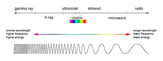
Figure 1: An illustration of the electromagnetic spectrum showing comparison between wavelength, frequency and energy. (Image Credit: NASA)
WHAT IS RADIATION?
There are two types of radiation: non-ionizing and ionizing, in this case meaning the ability to strip electrons from an atom. Non-ionizing radiation does not have enough energy to strip electrons from matter, thus there are no adverse health effects associated. Ionizing radiation, by contrast, does contain enough energy to strip electrons. X-rays are considered ionizing radiation. Ionization of essential cellular mechanisms, such as DNA strands and certain proteins, may cause cellular degradation and potentially cancerous mutations. However, when used properly with sufficient safety mechanisms, x-rays have many safe and useful applications. The following sections will detail how x-rays can be used to be more helpful than harmful to health and safety.
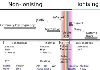
(Figure 2): The electromagnetic spectrum showing ionizing and non-ionizing radiation. (Image Credit: Wikipedia)
MEDICAL X-RAYS
Medical x-ray systems are used in a variety of exams so doctors can non-invasively and painlessly assess a patient’s health, diagnose diseases, and prepare for potential surgical procedures. X-ray scans may also help medical professionals in more practical applications such as the insertion of stents, catheters, or other necessary internal medical devices.
There are a few types of medical x-rays that use ionizing radiation:
Radiography: A single x-ray image recorded for later evaluation (e.g. dental x-rays).
Fluoroscopy: A continuous x-ray image displayed on a monitor. This allows for real-time monitoring of procedures or passage of contract agents (“dyes”) through the body.
Computed Tomography (CT): A detector moves around the patient’s body recording x-ray images. Computers then reconstruct the individual scans into cross-sectional images (“slices”) showing internal organs and tissues.
During these scan procedures, an x-ray beam passes through the patient’s body. Some of the x-rays are absorbed or scattered by the internal structures (bones, tissues, etc.). Detectors capture any remaining x-ray pattern, and computers process the pattern into images the doctor can review. The capabilities and radiation exposures involved in medical x-rays differ depending upon the type of scan used. Radiologists are trained to monitor the amount of x-rays that a patient is exposed to, and they ensure that patients are never exposed to potentially hazardous amounts.
CT scans (previously called CAT scans) expose patients to a greater amount of radiation than typical medical x-ray systems, though still within limits that minimize risk to the patient’s health. This is because the detailed slices can only be obtained through longer x-ray exposures. During the procedure, the patient lies on a table which slides into a cavity in the center of the scanner. The machine rotates around the patient, rapidly capturing images from all directions. A separate control room then processes the images and generates detailed slices for doctors to review. Despite the increased radiation exposure, CT scans are often the best method for detecting threats like cancer. Their detailed image slices allow doctors to confirm a tumor’s presence, size, and precise location. Doctors can also determine whether a tumor is affecting or connected to other nearby tissues.
Medical x-rays are a very powerful healthcare tool. They can provide important life-saving information. The images produced reveal internal structures hidden by skin and bones. They carry the risk of some radiation exposure, but the potential benefits outweigh those limited risks.
SECURITY X-RAYS
X-ray imaging security is ideal for searching items without the need to open or complete manual inspections. X-ray scanners produce quick, reliable, and detailed images showing the shape, type, and location of all items within a closed container. X-ray systems vary in size from scanners for small bags, to cargo pallets, to cars and trucks, and even trains! They serve numerous markets including law enforcement, transportation, critical infrastructure, aviation and the military. Finding the right x-ray security system can be a simple process. The end user simply needs to know what objects they will be scanning and what threats they are looking to identify.
There are two (2) common types of x-ray security scanners:
• Cabinet X-Ray Systems: Contain an x-ray tube installed within a shielded enclosure. The enclosure is made up of a material, usually lead, that prevents most of the x-ray radiation from escaping the cabinet.
• Personal Security Screening Systems: Two types of personal electronic screening products are used by TSA to screen people at airports: General Use X-Ray Screening Systems and Millimeter Wave Screening Systems
CABINET X-RAY SYSTEMS
Cabinet x-ray systems are common at security sites worldwide, from critical infrastructure sites like government buildings and courthouses to airports and aviation sites, as well as ports and border crossings. These systems can be deployed for screening baggage, air cargo, vehicles and much more. Cabinet x-ray systems make it possible to inspect items quickly without damaging the contents inside.
Astrophysics manufactures a wide range of cabinet x-ray systems, each tailored to a specific mission and operating environment. The XIS-6545™ is a small baggage scanner that fits perfectly alongside security checkpoints in an airport terminal, where space is limited and efficiency is absolutely critical.
Air cargo facilities, which regularly handle cargo pallets and large baggage of many shapes and sizes, need something larger and more powerful. The XIS-1818DV 200kV™ is a good compromise between size, weight, and power that can effectively scan most pallets. If such a facility needs more power for more thorough screening, the Multi-View CT provides a much more powerful x-ray source and a 3D model recreation program to allow for extremely detailed inspection.
Ports and borders are mostly concerned with quickly screening cars and cargo carrying trucks as they pass through the facility. For each of these, Astrophysics delivers the HXC-LaneScan™ and HXP lines. The first is a drive-through scanner that can effectively scan an entire car from roof to axle, and the latter is a truck scanner that can provide a detailed scan of an entire trailer and its contents. Both are effective at countering smuggling and trafficking of illicit goods.
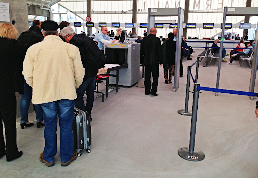
Figure 3: Cruise ship passengers line up at a port in Denmark for a routine security screening before boarding the cruise ship. (Image Credit: Astrophysics Inc.)
PERSONAL SECURITY SCREENING SYSTEMS
The first type of personal security screenings systems are those known as “General Use Systems”. These systems are also known as “backscatter” systems because they emit a very small amount of x-rays and measure the reflected radiation instead of the penetrating radiation. The reflected x-rays allow a detailed map of the surface of the scanned object, a person in this case.

Figure 4: A TSA officer screens a woman with a millimeter wave machine at an airport security checkpoint. (Image Credit: Reuters)
The second type of personal security screening systems are known as “Millimeter Wave systems”. Millimeter Wave systems use two different methods based upon the detection of millimeter waves which are non-ionizing and sit at the very high end of the radio wave band. Active systems emit radiation and measure the reflected patterns. By contrast, Passive systems detect the naturally occurring millimeter wave radiation emitted from most objects and the human body.
In today’s world it is common to screen items and people before allowing them access to a secure area. Airports, courthouses, government buildings, secure locations, cargo facilities, ports, border checkpoints, and more are all prime examples of locations requiring hardened security. Scanning technology is key to creating a more secure environment that enhances the capabilities of inspection personnel through advanced tools.
WHAT ARE THE DIFFERENCES BETWEEN SECURITY AND MEDICAL X-RAY SCANNERS?
Medical x-ray and security scanners are quite similar, with only a few key differences. Both technologies rely on the same fundamental concepts, the detection and interpretation of attenuated and/or reflected EM radiation. Their differences mostly center on the potential risks, required training, and methods of construction.
Medical x-ray scanners emit a lot of radiation, but must also adhere to strict standards because they directly irradiate human bodies. Medical professionals need specialized x-ray training to operate these machines and interpret the results. Training a radiologist is a long process spanning years of time, and requires years more of medical training to review and analyze scans.
Security scanning systems, by contrast, require an entirely different set of skills. While medical screening requires extensive medical training and an eye for details in monochrome images, security screening tests an inspectors ability to use a wide set of tools to quickly find and identify myriad different threats. Bags, cars, trucks, cargo pallets, and crates are all going to appear differently and may contain almost anything one could imagine; Inspectors must be able to very rapidly identify what an image contains and then spot threatening objects that may be hidden within, often with less than a minute per image. Systems include many manipulation tools and automated features to aid inspectors in this task, such as Astrophysics Enhanced Imaging™ (EI™) and AI™ software technology. Additionally, when scanning hundreds or thousands of items every day, x-ray security screening must be efficient and quick to process. The speed needed in security environments necessitates automation to maintain continuous operations.
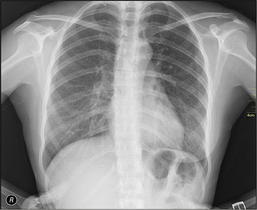
Figure 5: An example of a medical x-ray in black and white. (Image Credit: Nevit Dilmen/Wikipedia)
X-rays are not part of the visible spectrum, so they cannot have a “color” interpretable by humans. All x-ray images are produced in black and white. In security environments, systems employ coloration programs that categorize materials by type from organic materials (plastics, food, etc.) to heavy metals (steel, gold, lead, etc.) and assign each to a color. This is especially important because shape and density alone cannot determine what an object is. Inspection personnel can use both the original black and white image and the colored image to help analyze a scan.
Medical x-ray systems do not employ these kinds of coloration programs due primarily to the composition of the human body. Every part of the body is some kind of organic material, making coloration useless. In addition, coloration can sometimes hide details more clearly visible in black and white, making the inclusion of a coloration program unnecessary.
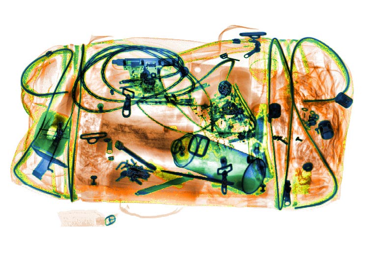
Figure 6: Colorized EI™ x-ray image of a duffel bag put through an Astrophysics x-ray scanner. The image assigns colors to scanned objects based on the objects’ atomic numbers. (Image Credit: Astrophysics Inc.)
ABOUT ASTROPHYSICS
Astrophysics is an x-ray security manufacturer with over 20 years of industry experience. With over 40,000 systems deployed in more than 150 countries, Astrophysics safeguards critical infrastructure, air cargo, ports, and border sites worldwide. Headquartered outside of Los Angeles, California, Astrophysics products are designed in the USA and assembled in North America to the highest quality standards. Astrophysics is also an International Organization of Standardization (ISO) 9001 and ISO 14001 certified organization. Our x-ray scanners adhere to rigorous industry specifications, and we offer TSA- and ECAC-qualified scanners for any security environment. Our superior imaging technology, relentless innovation, and customer service excellence have made Astrophysics the global brand of choice for x-ray security. Follow us on Twitter, LinkedIn, Facebook and YouTube for the latest news and x-ray product updates.